


- Your Questions Answered

- News
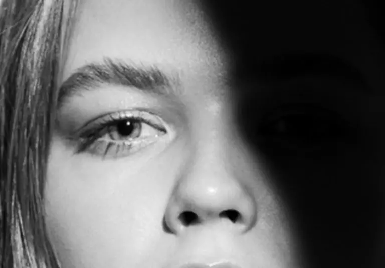
Keratoconus: Your Questions Answered
In this new LaserVision series, your questions answered, we aim to cover a number of pertinent topics, explaining any misconceptions, answering common questions and hopefully allaying any fears. This first edition covers keratoconus (Greek keras = cornea; latin conus=cone), a potentially visually disabling condition that is easily stopped if diagnosed and treated early.
What is it?
Keratoconus is a condition that causes your cornea, the clear front window of your eye, to weaken and change shape. It is thought that a biochemical and structural weakening of the cornea leads to this bowing and conical change in shape. It is surprisingly common and may in fact be under-reported due to delays in diagnosis. Untreated it can cause significant visual disability so regular reviews with a corneal specialist are essential.
What causes it and who is likely to get it?
It usually starts during the teenage years and can occur through to the early forties when it stabilises due to naturally occurring stiffening of the cornea which happens as we get older. Keratoconus is thought to be genetic and run-in families but in addition there are several other causes and associations (environmental factors such as eye rubbing / atopy or allergy; systemic conditions such as Marfan’s syndrome / Down syndrome) which are not fully understood. It may also occur randomly or sporadically.
What does it do to my vision? Does it change or get worse?
As the cornea becomes more cone shaped and distorted it may cause a progressive deterioration in vision. This may result in blurring, glare, haloes, or increased sensitivity to light. Unless treated, it is usually progressive and affects both eyes but to varying degrees.
How is it diagnosed?
It is diagnosed in clinic by a corneal specialist ophthalmologist. High astigmatism or progressive astigmatism is highly suspicious for the condition. The abnormal corneal shape is confirmed, and diagnosis made using a combination of clinical examination, glasses prescription (or refraction) and corneal tomography (advanced 3D mapping of the corneal shape and thickness).
What treatment is available?
The primary aim is to diagnose and treat as early as possible: stabilisation of the cornea is achieved using corneal collagen cross-linking (CXL) to prevent any further progression and change to vision. Riboflavin (vitamin B2) and UV-A light are applied to the corneal surface resulting in the formation of bonds between the collagen layers and overall stiffening of the cornea. Once stabilised, the treatment aim is to restore vision which can be achieved by glasses, contact lenses, intracorneal ring segments. It is also possible to combine CXL and laser to stabilise the keratoconus and smooth the corneal shape to improve the vision (Athens protocol). Sometimes in advanced disease, a corneal graft (DALK or penetrating keratoplasty) may be necessary to restore vision.
If you are concerned you or someone you know might have keratoconus or you have recently been diagnosed, please get in touch for a comprehensive review, assessment, and discussion of treatment options.
MEDICAL DISCLAIMER: this article is written as general information and should not be considered medical advice. It is always recommended to see your GP, ophthalmologist or other healthcare provider if any medical concerns.
Reviews
0% APR FINANCE
We offer 0% APR* finance for up to 48 months. To find out more information about which treatments are available with 0% APR finance, contact us today.
*subject to status
*minimum age applies
At LaserVision, we focus on you. Each consultation, treatment, procedure and aftercare plan is specifically tailored to your needs and requirements so you can rest assured you are getting the best treatment for you. Laser Vision Limited is authorised and regulated by the Financial Conduct Authority and is the broker and not the lender. Our FCA registration number is 805270. Laser Vision Limited offers credit products from Secure Trust Bank PLC trading as V12 Retail Finance. Credit is provided subject to affordability, age and status. Minimum spend applies. Not all products offered by Secure Trust Bank PLC are regulated by the Financial Conduct Authority
Please submit a compliment, concern, or complaint to: [email protected]. Every observation is an opportunity for our team to improve the quality of our patient care.
Sign up to our newsletter to receive updates
About
Our Patients
For Doctors
Office
LaserVision Ltd
Prema,
Compass Road
Portsmouth, PO6 4RP
United Kingdom
Contact Us
At LaserVision, we focus on you. Each consultation, treatment, procedure and aftercare plan is specifically tailored to your needs and requirements so you can rest assured you are getting the best treatment for you. Laser Vision Limited is authorised and regulated by the Financial Conduct Authority and is the broker and not the lender. Our FCA registration number is 805270. Laser Vision Limited offers credit products from Secure Trust Bank PLC trading as V12 Retail Finance. Credit is provided subject to affordability, age and status. Minimum spend applies. Not all products offered by Secure Trust Bank PLC are regulated by the Financial Conduct Authority
Please submit a compliment, concern, or complaint to: [email protected]. Every observation is an opportunity for our team to improve the quality of our patient care.
Sign up to our newsletter to receive updates
- © Copyright 2025
- Privacy
- Complaints
- Modern Slavery
- Sitemap
- Carbon Reduction Plan
![]() Site by Blow Media
Site by Blow Media